
Otoconia
Microscopic calcium-carbonate crystals that normally sit in the utricle. Patients often hear them called crystals, particles, rocks, or canaliths.
BPPV treatment is not one maneuver for every patient. We identify the involved ear, semicircular canal, and particle behavior, then use the eye-movement pattern to select and verify the appropriate repositioning maneuver.
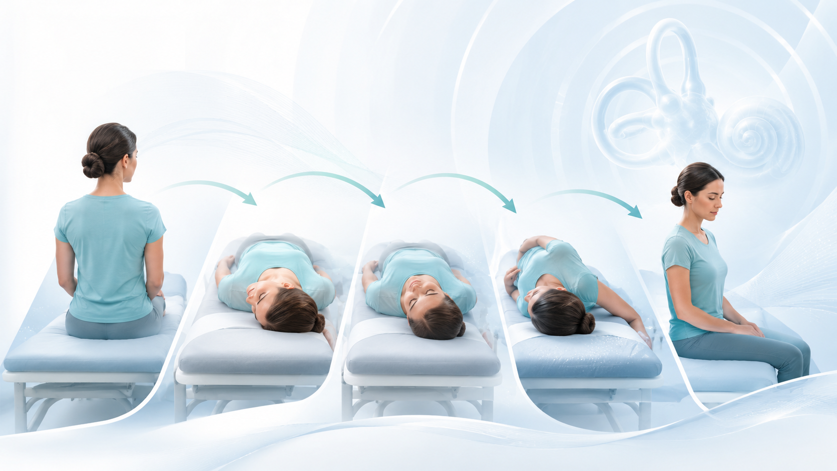
These videos are educational and are not a substitute for an examination. The wrong side or maneuver can fail, convert BPPV into another canal, or delay evaluation of a condition that is not BPPV. Do not attempt a home maneuver unless a qualified clinician has identified your pattern and confirmed that the movement is safe for your neck, back, circulation, and mobility.
BPPV is a mechanical inner-ear disorder, but the treatment plan is built from the history, positional testing, and nystagmus rather than symptoms alone.
Typical BPPV causes brief attacks of spinning or motion triggered by rolling in bed, lying back, looking up, bending, or rising.
Dix-Hallpike, side-lying, straight head-hanging, and supine roll tests are selected according to the suspected canal and the patient’s mobility.
Latency, direction, torsion, duration, fatigability, and reversal of nystagmus help localize the ear, canal, and mechanism.
The chosen maneuver is followed by repeat positional testing when appropriate. Persistent or changed nystagmus may require another maneuver or a revised diagnosis.
Microscopic calcium-carbonate crystals that normally sit in the utricle. Patients often hear them called crystals, particles, rocks, or canaliths.
Free-floating otoconia within a semicircular canal. Movement creates a transient fluid deflection, so the positional nystagmus typically has latency and fades.
Particles attached to or affecting the cupula. The nystagmus may begin quickly and persist while the provoking position is maintained.
Particles move from one canal into another during testing or treatment. A new nystagmus pattern calls for repeat localization, not automatic repetition of the first maneuver.
This is the language used to organize a positional examination. Nystagmus must be interpreted in the full clinical context, especially when it is persistent, purely vertical, atypical, or does not respond as expected.
| Suspected location | Typical provoking test | Typical observed pattern | Common treatment direction |
|---|---|---|---|
| Posterior canal | Dix-Hallpike or modified side-lying | Upbeating and torsional; the upper poles beat toward the involved ear in classic canalithiasis | Side-specific Epley/canalith repositioning; Semont-type liberatory maneuver in selected cases |
| Horizontal/lateral canal | Supine roll test | Direction-changing horizontal nystagmus: geotropic or apogeotropic. Relative intensity and secondary signs help determine the involved side. | Lempert/barbecue roll, Gufoni, Appiani, or another variant matched to the mechanism and side |
| Anterior/superior canal | Straight head-hanging and/or Dix-Hallpike | Downbeating with a torsional component may occur. Central and atypical posterior-canal patterns must also be considered. | Deep head-hanging/Yacovino or a side-specific reverse maneuver after careful localization |
Important: Geotropic does not simply mean “the side that feels worse,” and apogeotropic lateral-canal BPPV may represent more than one particle location. That is why the eye recording and response to each position matter.
The aim is to guide displaced otoconia through the involved canal and back toward the utricle. The sequence changes with anatomy and particle behavior.
The side-specific Epley maneuver is a standard first-line treatment. A Semont or modified maneuver may be selected for anatomy, mobility, cupular involvement, or treatment response.
Geotropic and apogeotropic variants are not treated identically. The clinic may use a Lempert roll, Gufoni, Appiani, Zuma, forced prolonged positioning, or a staged conversion strategy.
A deep head-hanging or Yacovino-type maneuver can be used when the findings support anterior-canal involvement. Downbeat positional nystagmus warrants careful neurologic interpretation.
Treatment is prioritized by the clearest or most symptomatic pattern, then repeated testing determines whether another canal or ear also requires treatment.
Use the sequence assigned by your clinician. Pause if you develop neck or back pain, fainting, new weakness or numbness, a severe headache, vision or speech change, or symptoms unlike your familiar positional vertigo.
A right-sided canalith repositioning sequence for a confirmed right posterior-canal pattern.
A left-sided canalith repositioning sequence for a confirmed left posterior-canal pattern.
A right-sided lateral-canal repositioning sequence selected after supine roll testing.
A left-sided lateral-canal repositioning sequence selected after supine roll testing.
Severe symptoms, restricted mobility, multi-canal disease, difficult localization, repeated treatment failure, or canal conversion may call for computer-assisted testing and treatment. The Epley Omniax and TRV chairs allow controlled multi-axis positioning while nystagmus is observed or recorded.
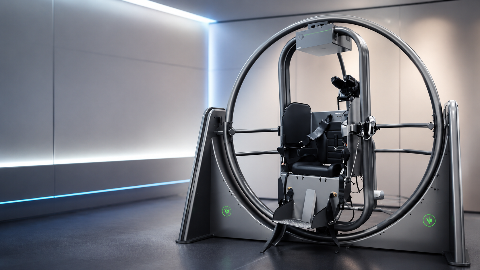
The provoking side alone is not enough. The diagnosis comes from the direction and intensity of nystagmus during canal-specific positional tests. Lateral-canal patterns are particularly easy to misidentify without observing both sides.
Particles may have moved without fully exiting, entered another canal, or revealed a second involved canal. The next step is repeat positional testing so the new pattern can direct treatment.
Yes. Recurrence does not necessarily mean the first treatment failed. A new episode may involve the same ear and canal or a different pattern, so relocalization is important before repeating a maneuver.
Residual dizziness can persist after successful repositioning. The clinician may recheck for remaining BPPV and consider vestibular rehabilitation, migraine, neuropathy, medication, cardiovascular, or neurologic contributors when symptoms continue.
No. Home treatment is most useful after the diagnosis, side, sequence, and safety considerations are established. A history of significant neck or back disease, vascular concerns, retinal problems, recent surgery, severe mobility limitation, or an atypical examination deserves clinician guidance.
Educational content only. Diagnosis and treatment must be individualized by a qualified clinician.
We evaluate the eye-movement pattern, localize the involved canal, and build a treatment plan for straightforward, recurrent, and complex positional vertigo.